| First Section Page | Page 6 of 7 | Last Section Page |
Axillary Access
(continued)
Step 5: Advance the wire through the needle and into the vein approximately 15-20cm. If the wire will not thread, place your thumb on the skin below the needle and retract the skin. Then, gently thread the wire. Wire can be cut on bevel of needle; be careful not to retract the wire through the needle. Maintain visibility and control of the wire at all times.
Step 6: Remove the access needle by sliding it out of the skin along the wire. Use more Lidocaine, if desired, prior to inserting the introducer. Perform skin nick if necessary. Slide the dilator/introducer over the wire.
Step 7: Slide the introducer along the wire track, using a twisting motion as needed, insert the introducer all the way into the vein. Remove the dilator and stabilize wire. Slowly thread the catheter over the wire. The wire will exit the distal lumen. Grasp the wire, maintaining control. Gently advance the catheter to premeasured internal length. Remove the wire and immediately place thumb over hole. Withdraw to confirm placement within vessel. Flush well with saline.
Step 8: Place needleless connector on each catheter lumen. Clean the insertion site to prepare for dressing and securement. Secure catheter using sutures or a manufactured securement device. Apply sterile dressing per facility policy.
Step 9: Confirm catheter terminal tip placement in the superior vena cava (SVC) using x-ray, fluoroscopy or electrocardiogram (EKG) per facility policy.
Femoral Access
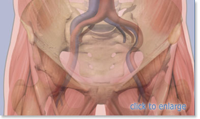
Femoral access is used as a last resort due to the high risk of infection and is used only in emergent situations. Once femoral access is obtained, perform evaluation every 24 hours as necessary to establish another lower risk device based on medical condition of the patient. Ultrasound should be used for scanning and needle-guided access to the femoral vein. Consider using a double prepping site procedure to counteract the high risk of infection. If possible, have the patient bathed with Chlorhexidine wipes prior to procedure. The femoral vein is usually located medial to the femoral artery.
Step 1: After performing a pre-scan and marking the insertion site, place patient in supine position. Perform hand hygiene and establish sterile field. Prep insertion site using Chlorhexidine. Allow to dry. Then, drape patient using maximal sterile barrier precautions.
| |
Page 6 of 7 |