| First Section Page | Page 5 of 7 | Last Section Page |
Axillary Access
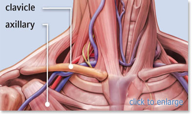
Insertion into the axillary vein is preferred because it allows you to do a modified subclavian insertion with visual aide of ultrasound. This region has a lower risk of pneumothorax or subclavian artery puncture. The axillary vein is located at the lateral border of the upper chest just past the shoulder. Pre-scanning with ultrasound is recommended to isolate the axillary vein, measure to ensure proper catheter length and vessel patency. In most cases, a 20cm catheter will be required.
Step 1: After performing a pre-scan and marking the insertion site, place patient in supine position. Perform hand hygiene and establish sterile field. Prep insertion site using Chlorhexidine. Allow to dry. Then, drape patient using maximal sterile barrier precautions.
Step 2: Scan the marked area to locate vein. Administer Lidocaine.
Step 3: Insert needle with syringe attached using a low angle of insertion with longitudinal access (25-30 degrees) or a higher angle with transverse access (60-80 degree). Image on screen should show dimpling of the vein as the needle touches it, then the vein rebounding as needle penetrates vein. Aspirate for blood return.
Step 4: Once aspiration of blood is confirmed, hold needle steady. Disconnect syringe and immediately place finger over hub of needle to prevent entrance of air.
| |
Page 5 of 7 |